[最も選択された] e coli under microscope 223876-How does e coli look under a microscope
Place your prepared slides next to each other on a paper towel or staining rack Cover the slides in the chosen stain and leave for 12 minutes Remove excess stain by running a gentle stream of water over the surface of the glass slide Place the glass slide on the microscope stage and begin viewing the specimenFind the perfect E Coli Under A Microscope stock photos and editorial news pictures from Getty Images Select from premium E Coli Under A Microscope of the highest qualityPrepared microscope slide of Escherichia coli, bacilli, smear Description This slide of a smear of E coli is best viewed under a microscope with an oilimmersion objective Qty 1 slide

Microscope Imaging Of Methylene Blue Stained E Coli Cells Harbouring Download Scientific Diagram
How does e coli look under a microscope
How does e coli look under a microscope-Get a second escherichia coli bacteria (e coli) stock footage at 2997fps 4K and HD video ready for any NLE immediately Choose from a wide range of similar scenes Video clip id Download footage now!Escherichia coli and Staph aureus ()jpg 2,160 × 1,440;
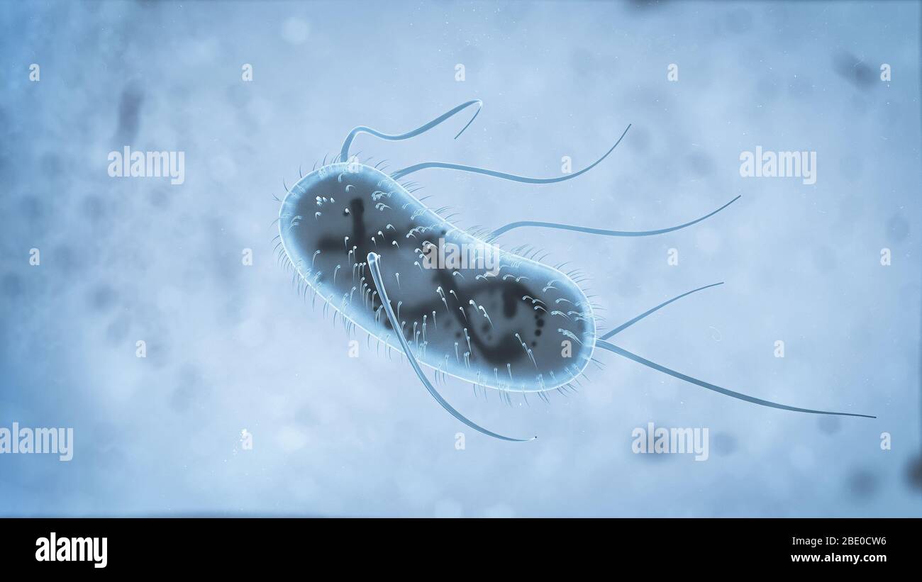


3d Escherichia Coli E Coli Cells Or Bacteria Under Microscope 3d Illustration Stock Photo Alamy
Which bacteria look similar to E coli under 100X optical microscope?Many bacteria look like E coli when examined under the microscope (if not stained Enterobacteriaceae, Bacillus, cornyeformeE coli Lform cells grew under aerobic conditions and required FtsZ (JoseleauPetit et al, 07), whereas peptidoglycan synthesis was required for their conversion and/or proliferation (JoseleauPetit et al, 07;
Ecoli bacteria growing in dish e coli under a microscope stock pictures, royaltyfree photos & images Bacteria culture that shows a positive infection of enterohemorrhagic E Coli, also known as the EHEC bacteria, in a patient lies on a table in theEscherichia coli, often abbreviated E coli, are rodshaped bacteria that tend to occur individually and in large clumps E coli are classified as facultative anaerobes, which means that they grow best when oxygen is present but are able to switch to nonoxygendependent chemical processes in the absence of oxygenAbout 15pm B What organelles are present in E coli?
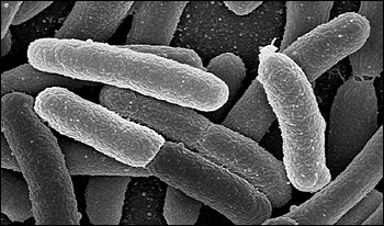
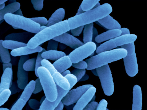
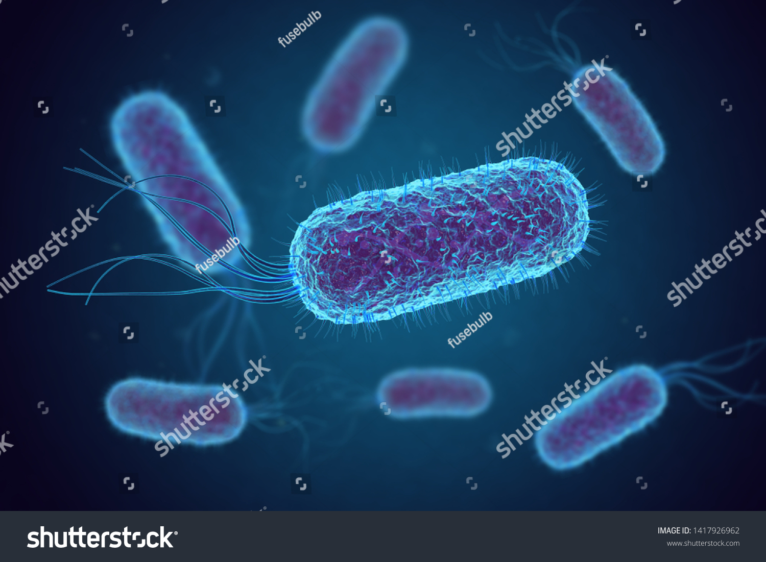
A picture of E coli under a microscope is shown above The tiny, hairlike structures you see help the bacteria attach itself to the surfaces inside the body, such as the liningE coli under the microscope Escherichia coli (E coli) is a bacterium commonly found in various ecosystems like land and water Most of the strains of E coli are harmless, but some strains are known to cause diarrhea and even UTIsEscherichia coli is shaped in the form of a rod It is a gramnegative bacterium and it contains peptidoglycan which is a thin and outer membrane Ecoli is considered a facultative thermophile, which means that it can survive under high temperatures as well as moderate temperatures It is present in most mammals particularly in the gut where it performs important functions like aiding in


Microscope Imaging Of Methylene Blue Stained E Coli Cells Harbouring Download Scientific Diagram


Gram Stain Wikipedia
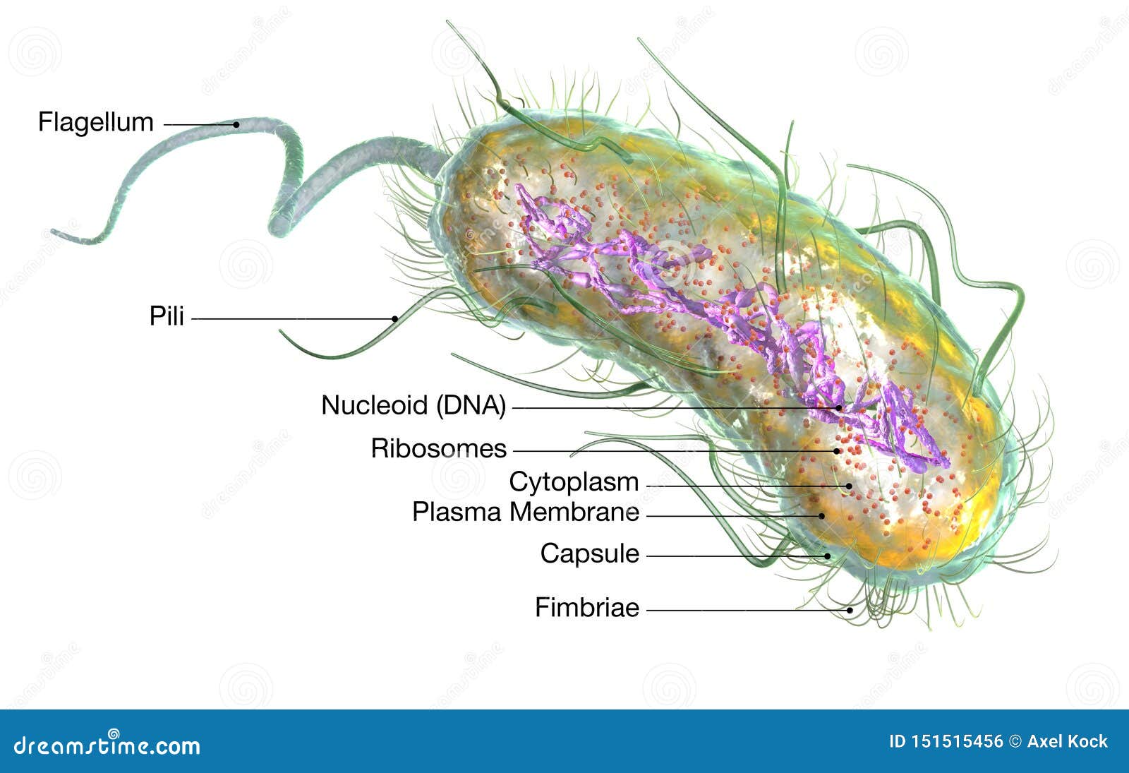
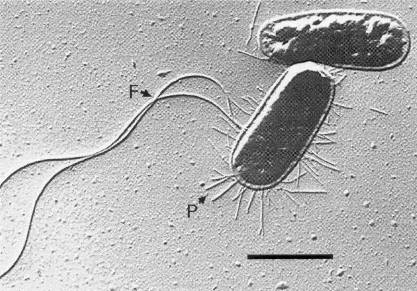
In the present study, the synergetic effect and mechanism of ultrasound (US) and slightly acidic electrolyzed water (SAEW) on the inactivation of Escherichia coli ( E coli ) were evaluated The results showed that US combined with SAEW treatment showed higher sanitizing efficacy for reducing E coli than US and SAEW alone treatmentEcoli is usually motile in liquid or semisolid environment with peritrichous flagella (about 6 per cell) and its surface is covered with fimbriae These structures (flagella and fimbriae) are too thin to be visualized by classical light microscopy or they don't have to be present at all under given cultivation conditions even at motile strains4 Observe Click on the cow and observe E coli under the highest magnification Notice the microscope magnification is larger for this organism, and notice the scale bar is smaller A What is the approximate size of E coli?


Escherichia Coli Slide W M Science Lab Microbiology Supplies Amazon Com Industrial Scientific


Image Of The Month E Coli Bacteria Growing On Mini Guts
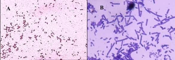
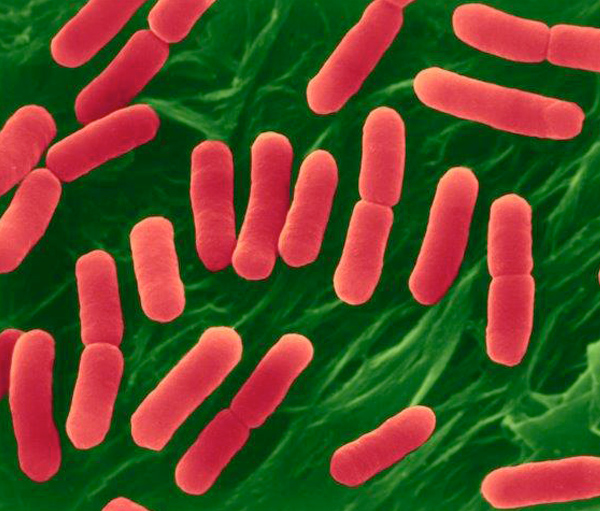
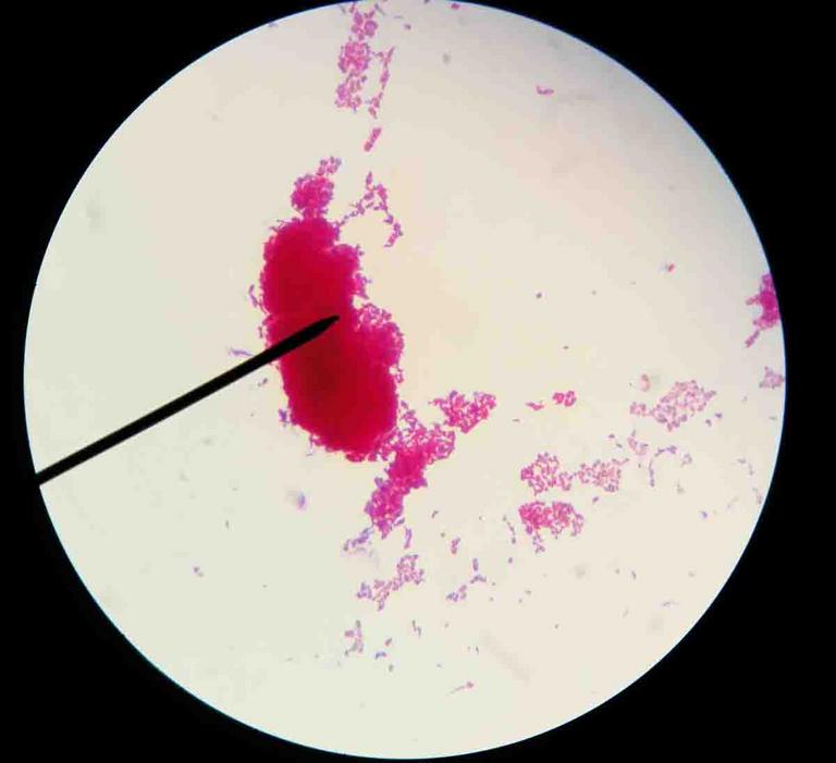
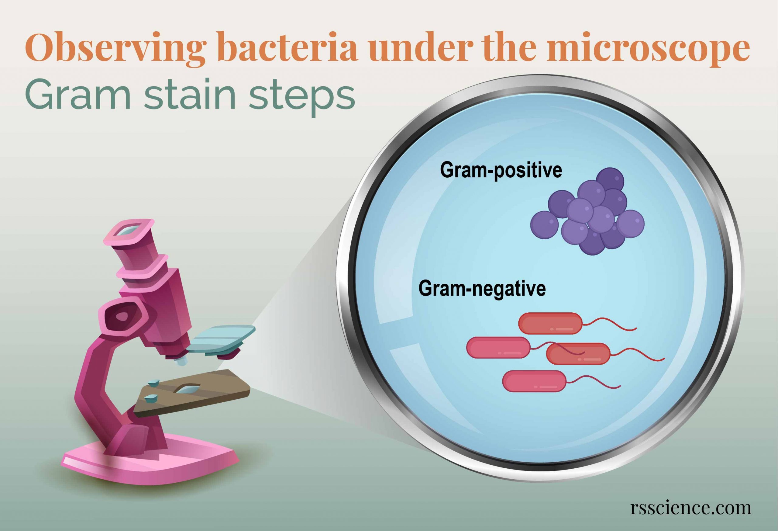
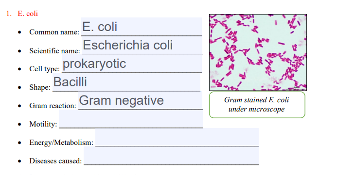
E coli bacteria looks like small rods under compound microscope at 1000 magnification power It is gram negative bacterium which appears pink when stained by a staining technique called as Gram staining which is a differential staining technique 942 viewsE coli is one of the key prokaryotic model organisms used in the fields of biotechnology and microbiology Hence, in many recombinant DNA experiments, E coli serves as the host organism The reasons behind using E coli as the primary model organism are some characteristics of E coli such as fast growth, availability of cheap culture media to grow, easiness to manipulate, extensiveEscherichia is a gramnegative bacterium, which under the microscope is shaped like a rod with a small tail It is widely distributed in nature (Brooker 08) Escherichia coli (E coli) is part of the normal intestinal flora Some strains are pathogenic and can cause gastroenteritis, UTI, meningitis, and wound infections
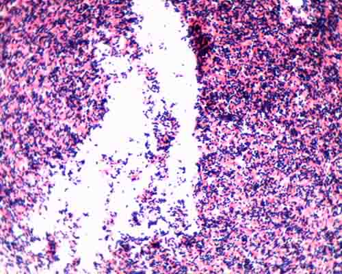


Gram Stain Microbiology Images Photographs


Q Tbn And9gctqlwezc G Rsexb5gmw Uv65za98k6p92dhlvblkp4mnrofyo Usqp Cau
Researcher with e coli bacteria bacteria under microscope stock pictures, royaltyfree photos & images ebola outbreak infographic bacteria under microscope stock illustrations chemical test of water bacteria under microscope stock pictures, royaltyfree photos & imagesStaphylococcus aureus and Escherichia coli under microscope, morphology and microscopic appearance Bacteria under Microscope Staphylococcus epidermidis Bacteria under Microscope Gramstain Grampositive Microscopic appearance Cocci in grapelike clusters, diplococci, cocciThe virulent E coli bacterium that killed dozens of people this spring will shortly land under the microscope as part of a European project to better understand dangerous pathogens


Lab 1


Gram Positive And Gram Negative Rods Microscopy Microbiology Microbiology Lab
The Best E Coli Microscope of 21 – Reviewed and Top Rated After hours researching and comparing all models on the market, we find out the Best E Coli Microscope of 21 Check our ranking below 2,9 Reviews Scanned2 Micrococcus luteus or Staphylococcus epidermidis a Fix a smear of either Micrococcus luteus or Staphylococcus epidermidis to the slide as follows 1First place a small piece of tape at one end of the slide and label it with the name of the bacterium you will be placing on that slide 2Using the dropper bottle of deionized water found in the staining rack, place 1/2 of a normal sizedE coli under the microscope Escherichia coli (E coli) is a bacterium commonly found in various ecosystems like land and water Most of the strains of E coli are harmless, but some strains are known to cause diarrhea and even UTIs E coli is commonly studied as they are considered as a standard for the study of different bacteria


Staphylococcus Aureus And Ecoli Under Microscope Microscopy Of Gram Positive Cocci And Gram Negative Bacilli Morphology And Microscopic Appearance Of Staphylococcus Aureus And E Coli S Aureus Gram Stain And Colony Morphology On Agar Clinical


Ecoli Frequently Asked Questions Escherichia Coli What Is Ecoli
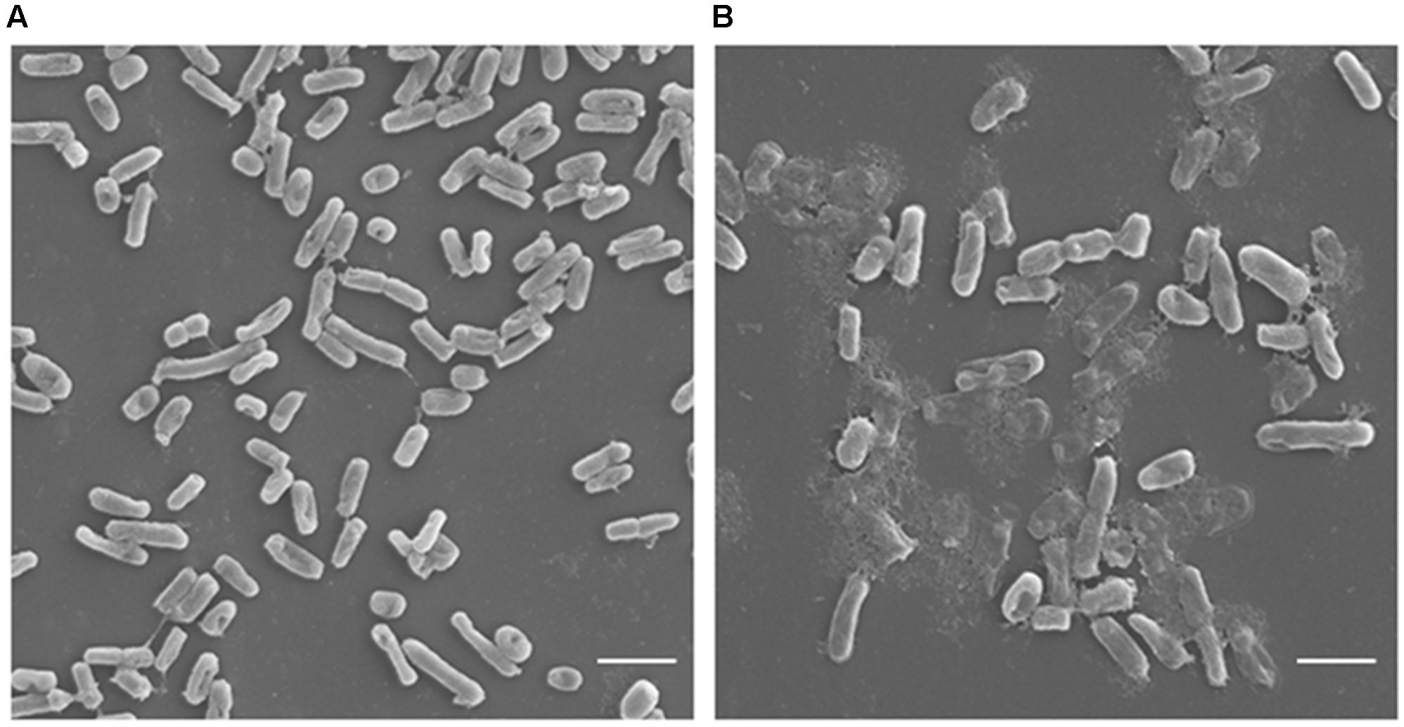
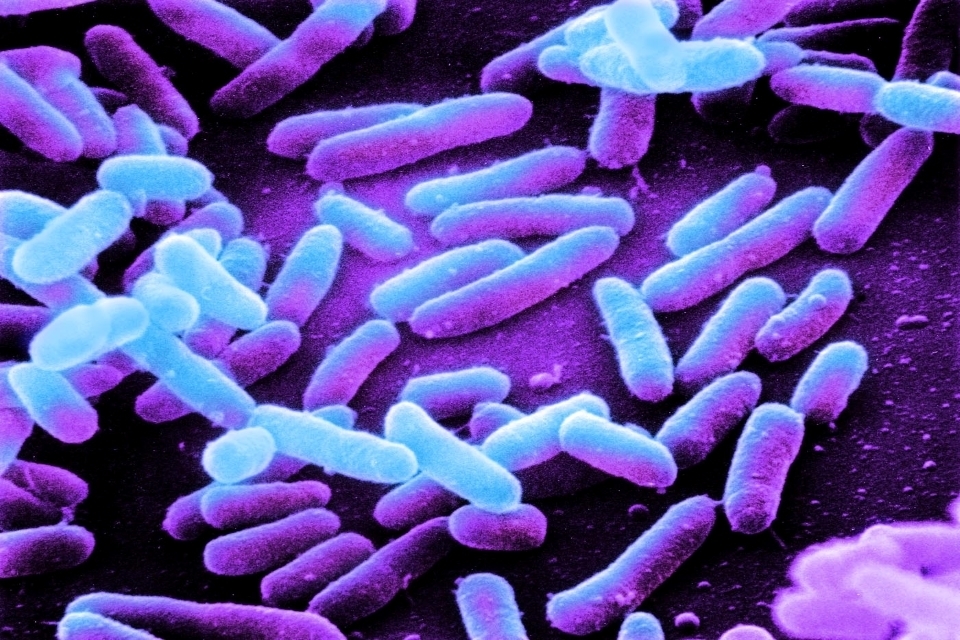
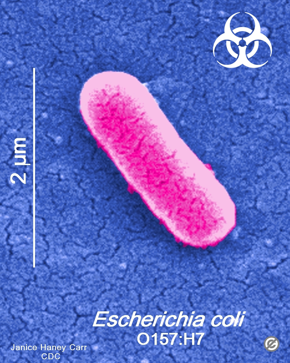
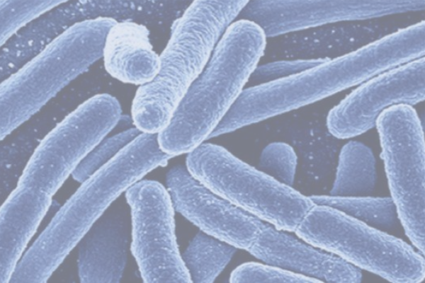
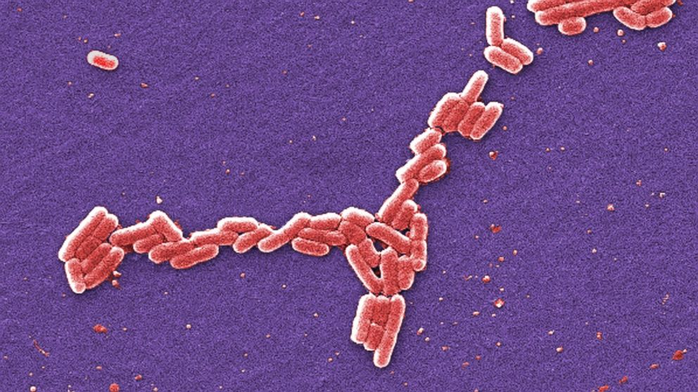
Under a high magnification of 66X, this digitallycolorized, scanning electron microscopic (SEM) image depicted a growing cluster of Gramnegative, rodshaped, Escherichia coli bacteria, of the strain O157H7, which is a pathogenic strain of E coliObservation When viewed under thePlace the slide on a staining rack and add a few drops of crystal violet onto the sample, gently wash with water Add a few drops of Gram iodine (mordant) for between 30 seconds and 1 minute, gently wash with water Add a few drops of alcohol (95% alcohol) for about 10 seconds, gently wash with water
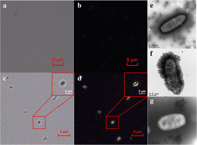


Ultrasensitive And Rapid Count Of Escherichia Coli Using Magnetic Nanoparticle Probe Under Dark Field Microscope Bmc Microbiology Full Text
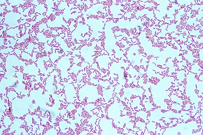


Cyber Fish Staphylococcus Aureus Or E Coli Neisseria
In liquid culture media like Trypticase soy broth or Nutrient broth, the growth of the bacterium occurs as a turbidity in the broth medium with a heavy deposits that disperses in the medium on shaking, which is further analyzed for the morphology (under the microscope), gram reaction, biochemical tests, and Escherichia coli specific tests In Blood Agar medium, some of the strains show betaE Coli under a microscope This is the one that can make you seriously ill and is responsible for the majority of Ecoli cases If you love traveling you may have already met Ecoli, it's commonly found when traveling to less developed countries and results in diarrheaE Coli E Coli is a more common bacterium than Salmonella which causes serious food poisoning in food Its scientific name is gramnegative bacteria The red shaped E Coli is facultative anaerobic bacterium while its scientific notion is presented as Escherichia coli or E coli These types of bacteria prefer living in warm blooded animals
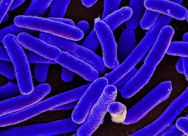


E Coli Under The Microscope Types Techniques Gram Stain Hanging Drop Method


All You Need To Know About E Coli Food Poison Journal
• A basic light microscope has 4 objective lenses – 4X, 10X, 40X, and 100X The higher the number, the higher the power of magnification Once you have focused theWhen viewed under the microscope, Gramnegative E Coli will appear pink in color The absence of this (of purple color) is indicative of Grampositive bacteria and the absence of Gramnegative E ColiUropathogenic Escherichia coli strains isolated from these patients, as well as the archetypal E coli strain UTI, were found to produce colibactin In a murine model of UTI, the machinery producing colibactin was expressed during the early hours of the infection, when intracellular bacterial communities form


Escherichia Coli Colony Morphology And Microscopic Appearance Basic Characteristic And Tests For Identification Of E Coli Bacteria Images Of Escherichia Coli Antibiotic Treatment Of E Coli Infections
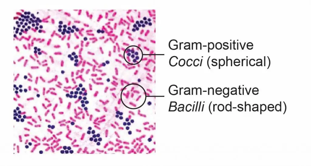


Observing Bacteria Under The Microscope Gram Stain Steps Rs Science
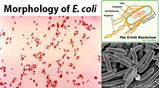
Ecoli is usually motile in liquid or semisolid environment with peritrichous flagella (about 6 per cell) and its surface is covered with fimbriae These structures (flagella and fimbriae) are too thin to be visualized by classical light microscopy or they don't have to be present at all under given cultivation conditions even at motile strainsEscherichia coli (E coli) bacteria normally live in the intestines of people and animals Most E coli are harmless and actually are an important part of a healthy human intestinal tract However, some E coli are pathogenic, meaning they can cause illness, either diarrhea or illness outside of the intestinal tract The types of E coli that can cause diarrhea can be transmitted throughCell wall, cell membrane, cytoplasm, flagellum, pilus, and nucleoid C What organelle is missing from E coli?
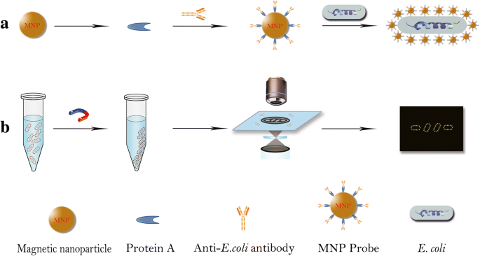


Ultrasensitive And Rapid Count Of Escherichia Coli Using Magnetic Nanoparticle Probe Under Dark Field Microscope Bmc Microbiology Full Text
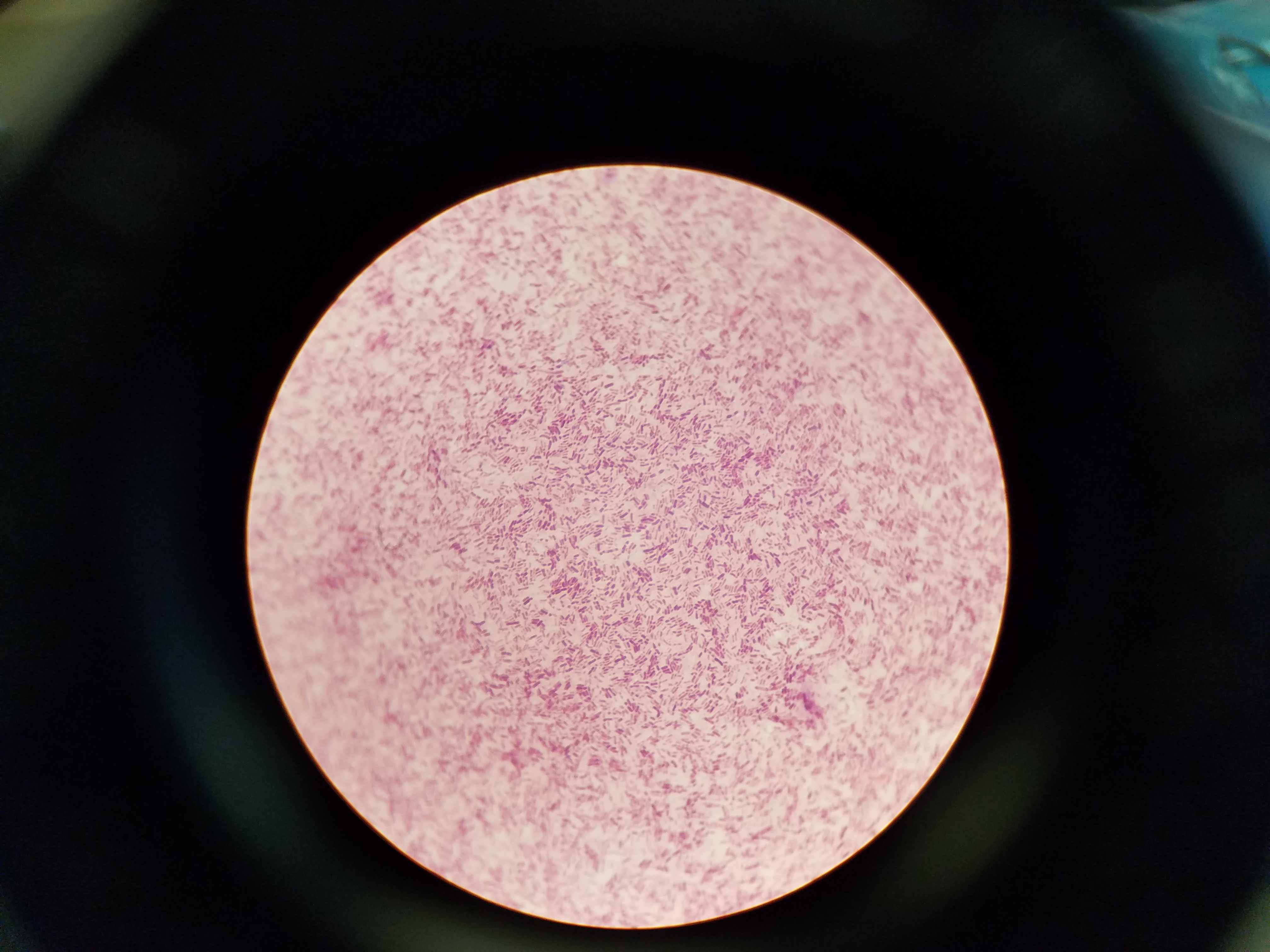


File Microbiology Gram Stain Jpg Wikimedia Commons
Escherichia is a gramnegative bacterium, which under the microscope is shaped like a rod with a small tail It is widely distributed in nature (Brooker 08) Escherichia coli (E coli) is part of the normal intestinal flora Some strains are pathogenic and can cause gastroenteritis, UTI, meningitis, and wound infectionsE coli 1 Escherichia coli is a Gram negative, facultative anaerobic, rodshaped bacteria It is a commensal that is found inhabiting the lower intestine of warm blooded animals A small proportion of E coli strains are pathogenic The harmless strains produce vitamin K and prevent colonization of the intestine by pathogenic bacteria E coli is classified into serotypes based on cell wallEscherichia coli, often abbreviated E coli, are rodshaped bacteria that tend to occur individually and in large clumps E coli are classified as facultative anaerobes, which means that they grow best when oxygen is present but are able to switch to nonoxygendependent chemical processes in the absence of oxygen


In Vitro Study Of The Activity Of Some Medicinal Plant Leaf Extracts On Urinary Tract Infection Causing Bacterial Pathogens Isolated From Indigenous People Of Bolangir District Odisha India Biorxiv


Q Tbn And9gcs3nlev0tx1pfsddwm6y9ajujk4lxjzmg7ksdxctgnipv0l50c Usqp Cau
The genus Salmonella is closely related to Escherichia coli bacteria and is suggested to have diverged from the bacteria (E coli) about 150 million years ago As such, it has adapted and can be found in several niches in the environment View the slide under the microscope starting with lower power;Prepared microscope slide of Escherichia coli, bacilli, smear Description This slide of a smear of E coli is best viewed under a microscope with an oilimmersion objective Qty 1 slide479 KB Escherichia coli flagella TEMpng 600 × 6;


Q Tbn And9gcrxsv6valktvydnaiqm6 Yoim4dbn 9td0atqk2azvfczn73wcq Usqp Cau
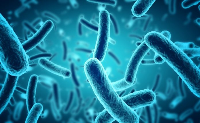


Particular E Coli Strain Can Cause Colorectal Cancer Finds Study The Federal
The length can range from 110 µm for filamentous or rodshaped bacteria The most wellknown bacteria E coli, their average size is ~15 µm in diameter and 26 µm in length In this figure The size comparison between our hair (~ 60 µm) and E coli (~1 µm) Notice how small the bacteria areCocci in grapelike clusters (Saureus) and bacilli(Ecoli) Clinical significance of Saureus Frequently found as part of the normal skin flora on the skin and nasal passages;Bacteria detected E coli, Staphylococcus, Streptococcus, Enterococcus, Campylobacter, Streptobacillus Cleaning contenders Cream cleaner vs Shower cleaner The shower head was the fourth dirtiest surface in the bathroom, with 32% covered in bacteria, but cream cleaner managed to reduce the infected area by a satisfying 99%


Zkfaa Bioproses Lab 1 Principles And Use Of Microscope


Introductory Chapter The Versatile Escherichia Coli Intechopen
E coli can spread to the urinary tract in a variety of ways Common ways include Your urine will then be examined under a microscope for the presence of bacteria Urine cultureMost of the bacteria range from 022 µm in diameter The length can range from 110 µm for filamentous or rodshaped bacteria The most wellknown bacteria E coli, their average size is ~15 µm in diameter and 26 µm in length In this figure The size comparison between our hair (~ 60 µm) and E coli (~1 µm) Notice how small theCells of Escherichia coli NBRC 3972 and Staphylococcus aureus NBRC were inoculated onto an agar (15%) medium varying in nutrient concentration from full strength of the nutrient broth (NB) to 1/10 NB Immediately thereafter, the inoculated agar was placed on antimicrobial and nonantimicrobial surfaces in such a way that the microbial cells came into contact with these surfaces


27 Earth Bacteria E Coli
.jpg)


Escherichia Coli 400x Escherichia Coli 400x Manufacturers Escherichia Coli 400x Suppliers Escherichia Coli 400x Exporters Escherichia Coli 400x In India
E coli is an example of a bacteria Bacteria are classified as prokaryotic cells from SCIENCE SNC2D at G A Wheable Secondary School Look at the Sand/silt sample under the microscope A Turn on Show labels Select the human skin sample On the MICROSCOPE tab, choose the 400x magnification, focus on the sample,Escherichia coli Escherichia coli is a small bacillus Estimate the length and width of a typical rod c Treponema pallidum Treponema pallidum is a spirochete a thin, flexible spiral On this slide you are examining a direct stain ofTreponema pallidum, the bacterium that causes syphilis Measure the length and width of a typical spirochete263 MB Escherichia coli electron microscopyjpg 800 × 640;


High Precision Characterization Of Individual E Coli Cell Morphology By Scanning Flow Cytometry Konokhova 13 Cytometry Part A Wiley Online Library


How These 26 Things Look Like Under The Microscope With Diagrams
Billings et al, 14) Contrasting evidence may be explained by different experimental conditions employed, such as aeration andIt is estimated that % of the human population are longterm carriers of S aureusEColi under the microscope FEATURED BOOK Microcosm E Coli and the New Science of Life WHAT DOES E COLI LOOK LIKE?


10pk Escherichia Coli Smear Gram Stain Prepared Microscope Slides 75 X 25mm Classroom Pack 10 Slides In Storage Case Biology Microscopy Eisco Labs Amazon Com Industrial Scientific


Frontiers Antimicrobial Potential Of Carvacrol Against Uropathogenic Escherichia Coli Via Membrane Disruption Depolarization And Reactive Oxygen Species Generation Microbiology
Escherichia coli Escherichia coli is a small bacillus Estimate the length and width of a typical rod c Treponema pallidum Treponema pallidum is a spirochete a thin, flexible spiral On this slide you are examining a direct stain ofTreponema pallidum, the bacterium that causes syphilis Measure the length and width of a typical spirocheteOur primary subject is the peritrichouslyflagellated bacterium Escherichia coli, that lives in your gut We are trying to learn how its flagellar motors work, how their directions of rotation are controlled by the cell's sensorytransduction network, and what effect that rotation has on modes of flagellar propulsionE Coli under the microscope at 400x


Escherichia Coli E Coli An Overview Microbe Notes


Bugs For Primary Schools Display Day
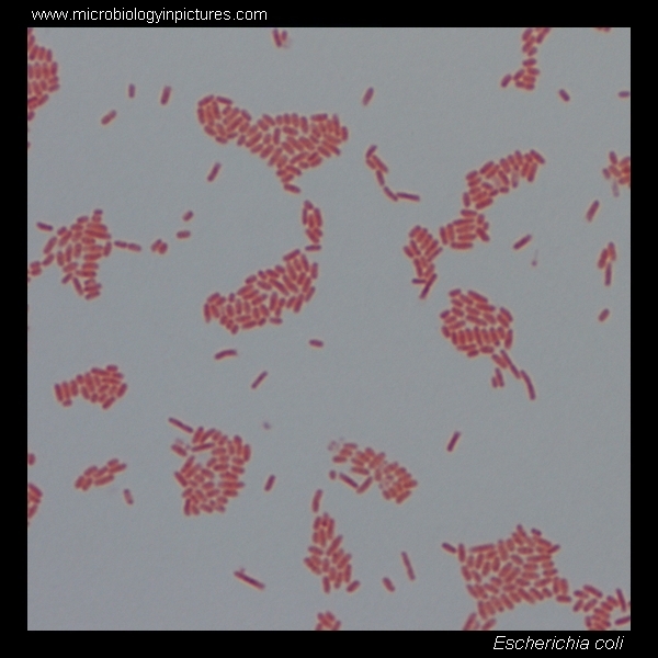


E Coli Gram Stain And Cell Morphology E Coli Micrograph Appearance Under The Microscope E Coli Cell Morphology E Coli Microscopic Picture


E Coli The Good The Bad And The 0157 By Pluralis Majestatis Medium


Escherichia Coli Wikipedia


641 E Coli Bacteria Illustrations Clip Art Istock


Escherichia Coli Wikipedia


Escherichia Coli E Coli National Geographic Society


Gram Stain Of E Coli Bacterium A Gram Stain Of Shows Gramnegative Download Scientific Diagram


19 3 Bacteria Videos And Hd Footage Getty Images
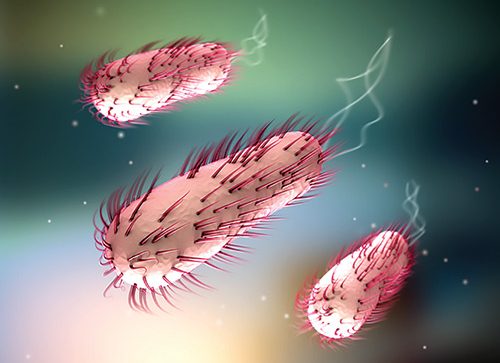


How E Coli Bacteria Launch Infections


Escherichia Coli Light Microscopy
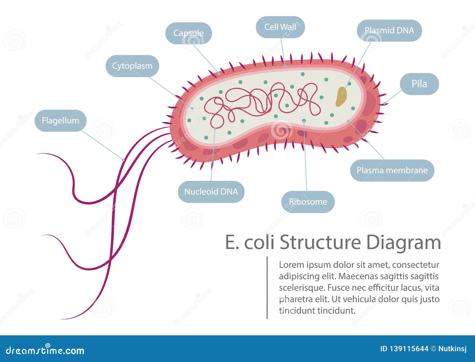


Escherichia Coli Structure Diagram Stock Vector Illustration Of Medical Diagram


4 490 E Coli Bacteria Stock Photos Pictures Royalty Free Images Istock


Graphy Bacteria Microorganism Coccus E Coli Microscope Technic Microscope Png Pngegg
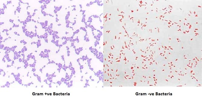


Gram Staining Principle Procedure Interpretation Examples And Animation


Staphylococcus Aureus And Ecoli Under Microscope Microscopy Of Gram Positive Cocci And Gram Negative Bacilli Morphology And Microscopic Appearance Of Staphylococcus Aureus And E Coli S Aureus Gram Stain And Colony Morphology On Agar Clinical


Biol 230 Lab Manual Gram Stain Of E Coli


E Coli Bacteria Abc News Australian Broadcasting Corporation


Optical Microscope Images Of E Coli Cells Following Gram Staining A Download Scientific Diagram


What Does An E Coli Bacteria Look Like Under A Microscope Quora


E Coli Escherichia Coli A Gram Negative Bacterium Causing Gastrointestinal Infection


Genomes Of Two Popular Research Strains Of E Coli Sequenced Bnl Newsroom


Escherichia Coli Bacteria E Coli Medically Accurate 3d Illustration Labeled Stock Illustration Illustration Of Biota Biology


Gram Negative Pink Colored Small Rod Shape E Coli Under Light Download Scientific Diagram


Escherichia Coli 07 Igem Org


E Coli O157 H7 Escherichia Coli Expert Witness And Epidemiology Services


Here S Why Drug Resistant Bacteria Could Spread Globally Time


Pathogenic E Coli


Acid Fast Stain Free Microbiology Images From Science Prof Online


3d Escherichia Coli E Coli Cells Or Bacteria Under Microscope 3d Illustration Stock Photo Alamy


Eiscoprepared Microscope Slide Escherichia Coli Smear Gram Stain Microbiology Fisher Scientific


E Coli Bacteria Light Microscopy Stock Video Clip K004 9042 Science Photo Library


Http Coltonanderson1 Weebly Com Uploads 2 4 3 0 Manual Pdf


Bacteria Under The Microscope E Coli And S Aureus Youtube


Phe Issues Advice To People Travelling To Egypt Gov Uk


Canada Food Recall Warning Unpasteurized Cider Recalled Due To E Coli O157 H7 Food Law Latest
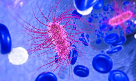


E Coli And Foodborne Illness Fda


E Coli Bacteria Microscopic Organism Indicates Fecal Contamination


Gram Stain Images Microbiology Stain Study Tools


Escherichia Coli Sem Stock Image C043 7660 Science Photo Library


E Coli Under The Microscope Types Techniques Gram Stain Hanging Drop Method


E Coli Under A Scanning Electron Microscope Image Eurekalert Science News
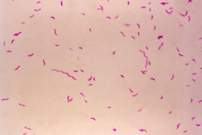


10 Foodborne Pathogens And Foodborne Illness Fight Bac


Difference Between E Coli And Klebsiella Lactose Fermentor Enterobacteriaece Youtube


2 194 E Coli Photos And Premium High Res Pictures Getty Images


Cell Division Of E Coli With Continuous Media Flow Youtube


Staining Microscopic Specimens Microbiology


Get The Facts About Hemorrhagic Colitis Caused By E Coli Everyday Health
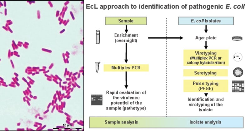


Escherichia Coli E Coli An Overview Microbe Notes


Optical Microscope Images Of E Coli Cells Following Gram Staining A Download Scientific Diagram


Escherichia Coli E Coli Cells Bacteria Stock Illustration


E Coli Why So Famous


Observing Bacteria Under The Microscope Gram Stain Steps Rs Science


Morphological View Of Lactococcus Culture Under Microscope 100x After Download Scientific Diagram


Escherichia Coli Bacteria E Coli Stock Footage Video 100 Royalty Free Shutterstock


Viruses And Bacteria Under Microscope 3d Rendered Bacteria And Viruses Stock Photo Alamy


Magnification Bioninja


Understanding E Coli In A Food Processing Context Fda Reader
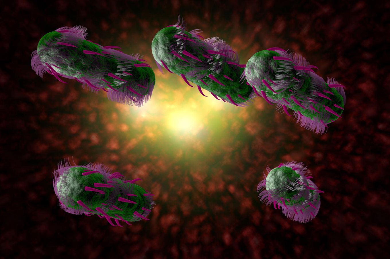


E Coli Food Poisoning
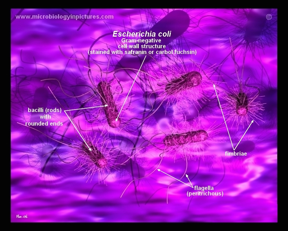


How E Coli Bacteria Look Like


Escherichia Coli Bacteria E Coli Stock Footage Video 100 Royalty Free Shutterstock


Escherichia Coli Bacteria E Coli Stock Footage Video 100 Royalty Free Shutterstock


Morphology Of E Coli Cells Under Microscope At 100 Magnification Download Scientific Diagram


Www Mccc Edu Hilkerd Documents Bio1lab3 Exp 4 Pdf


City In Oklahoma Ordered To Boil Water After Possible E Coli Contamination Abc News


Gram Stain


Understanding E Coli Symptoms Spread Prevention Cbc News


Escherichia Coli Bacteria E Coli Stock Footage Video 100 Royalty Free Shutterstock


Pathogenic E Coli


Gram Stain


A Long Chain Like Colony Of Streptococcus Bacteria As Seen Under Download Scientific Diagram


Solved 1 E Coli E Coli Common Name Escherichia Coli S Chegg Com


3


コメント
コメントを投稿